Year: 2012 (page 2 of 55)

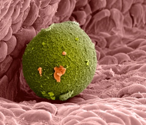
mapping human sex cells

Exciting studies on human reproductive cells are profiled in Nature:

Even though the reproductive age for humans is around 15–45 years old, the precursor cells that go on to produce human eggs or sperm are formed much earlier, when the fertilized egg grows into a tiny ball of cells in the mother’s womb. This ball of cells contains ‘pluripotent stem cells’ — blank slates that can be programmed into any type of cell in the body — and researchers are hoping to use these stem cells to treat various conditions, including infertility.


But little is known about the early developmental stages of human gametes — owing to the sensitivity of working with human tissue — and most work in this area has been conducted using mice. In a Nature Cell Biology paper today1, researchers from the University of California, Los Angeles, trace the development of early germ cells in human fetuses of between 6 to 20 weeks and analysed when genes were turned on or off.


The DNA within these early germ cells carries ‘epigenetic modifications’ — structural changes that do not affect the DNA sequence itself but do affect the way that genes are expressed. These changes may have accumulated during the parents’ lives, and need to be erased during the fetal stage. The study found two major events that wipe out, or reprogram, epigenetic modifications. Most of this reprogramming happened before 6 weeks, but the authors found a second event that completes the reprogramming after 6 weeks.

mistletoe

Mistletoe

The New York Times has an interesting piece on mistletoe’s role in nature. It’s a parasite but it appears to be key in maintaining a health and balanced forest ecosystem:

Dr. Watson, known in academic circles as “the mistletoe guy,” had long suspected that his favorite plant was a keystone species, meaning it punches above its weight, ecologically speaking, but even he was unprepared for the results. He had supposed that creatures that fed or nested on mistletoe would be affected by its removal. Instead, he found that the whole woodland community in the mistletoe-free forests declined.

Three years after the mistletoe vanished, so had more than a third of the bird species, including those that fed on insects. Bird diversity is considered an indicator of overall diversity. Where mistletoe remained, bird species increased slightly. It was a similar story for some mammals and reptiles, but, in another surprise, particularly for those that fed on insects on the forest floor.

“It’s a bit of a head-scratcher,” said Dr. Watson.

Analysis showed that species of mistletoe play an important role in moving nutrients around the forest food web. That has to do with their status as parasites.

Nonparasitic plants suck nutrients out of their own leaves before they let them fall, sending dry containers to the ground. But because the vampiric mistletoe draws water and nutrients from the tree stem or branch it attaches to, it is more nonchalant about leaving that nutrition in falling leaves. That means the fallen leaves still contain nutrients that feed creatures on the forest floor.

Not only that, but mistletoes make and drop leaves three or four times as rapidly as the trees they live off of, said Dr. Watson. As evergreens, they also do it throughout the year, even when trees are dormant. It is like a round-the-calendar mistletoe banquet.

platelets confer resistance to parasites

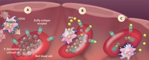
Platelets prevent parasitic infestation. Image from Sciencemag.org

From Science magazine this week, a report on how platelets express genes to help kill parasites. An excerpt from the perspective:

The six Plasmodium parasite species that cause disease in humans (P. falciparum, P. vivax, P. malariae, P. ovale wallickerie, P. ovale curtisii, and P. knowlesi) appear to have independently colonized hominids and influenced the genetic composition of different human populations (3). For example, the genes responsible for sickle cell anemia, thalassemia, and glucose-6-phosphate dehydrogenase deficiency have a higher frequency in populations where malaria is, or was once, endemic. These genes provide protection against severe malaria syndromes and likely evolved in response to the disease by providing a survival advantage (4). Another of these genes encodes the Duffy-antigen receptor for chemokines (DARC/Fy glycoprotein/CD234) found on red blood cells. This protein also acts as a receptor for P. vivax (5), and human red blood cells lacking this receptor are resistant to invasion by this species and by P. knowlesi (67). The impact of this genetic selection can be seen in the geographical distribution of P. vivax. This parasite is spread throughout tropical regions of the world, but is rare in large areas of central and western Africa where many individuals lack Duffy-antigen receptor expression on red blood cells. Thus, this “Duffy-negative” phenotype appears to have evolved as an innate resistance mechanism to P. vivax infection.

McMorran et al. extend previous work that demonstrated an important role for platelets in resistance to malaria (8) by identifying platelet factor 4 (PF4) as a key molecule in platelet-mediated killing of P. falciparum. PF4 is released from α granules in activated platelets to promote blood coagulation (9). It binds the Duffy-antigen receptor, along with several other chemokines (10). McMorran et al. found that a functional Duffy-antigen receptor is required for the antiparasitic activity of PF4.

Also check out the scientific research paper here [subscription required].

//chamoolroungas.net/4/4535925