Category: Chemistry (page 5 of 15)

dopamine explained


Dopamine

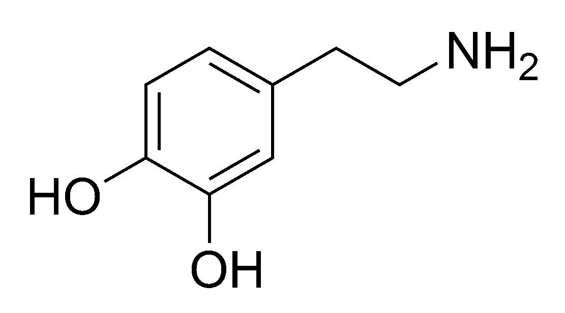
The structure of dopamine

Last week on Slate, Bethany Brookshire explains the neurotransmitter dopamine and how it effects many different chemical processes:

What is dopamine? Dopamine is one of the chemical signals that pass information from one neuron to the next in the tiny spaces between them. When it is released from the first neuron, it floats into the space (the synapse) between the two neurons, and it bumps against receptors for it on the other side that then send a signal down the receiving neuron. That sounds very simple, but when you scale it up from a single pair of neurons to the vast networks in your brain, it quickly becomes complex. The effects of dopamine release depend on where it’s coming from, where the receiving neurons are going and what type of neurons they are, what receptors are binding the dopamine (there are five known types), and what role both the releasing and receiving neurons are playing.

It has far more roles in the brain to play. For example, dopamine plays a big role in starting movement, and the destruction of dopamine neurons in an area of the brain called the substantia nigra is what produces the symptoms of Parkinson’s disease. Dopamine also plays an important role as a hormone, inhibiting prolactin to stop the release of breast milk. Back in the mesolimbic pathway, dopamine can play a role in psychosis, and many antipsychotics for treatment of schizophrenia target dopamine. Dopamine is involved in the frontal cortex in executive functions like attention. In the rest of the body, dopamine is involved in nausea, in kidney function, and in heart function.
the see through brain

A mouse brain imaged using the CLARITY technique.

Brain research has been getting lots of press since President Barack Obama announced his BRAIN initiative to map the human brain. Earlier this week, Nature published a paper by a group of Stanford researchers or a new technique called CLARITY. This technique allows the researchers to make whole organs transparent. They can then use different chemical compounds to label and highlight specific cells or features of the organ. They were able to demonstrate on brain tissue. From Popular Science:

Making these images is an eight-day process. The Stanford researchers started by infusing a mouse brain with a hydrogel solution. They then put the gel and brain into an incubator to set. (Like making Jell-O! Except that the setting, in this case, required a higher temperature rather than a lower one.)

The set gel bound to and physically supported most of the things in the brain. The gel didn’t bind to lipids, or fats, in the brain, however. Such fat is opaque and surrounds each cell. When researchers extracted this unbound fat, they were left with a clear view of everything else, frozen in place. For example, proteins that were originally embedded in cell membranes and the little spines that come off of neurons both remained.

At this point, the researchers could add different molecules to color the parts of the brain they want to study and look at the whole thing under a light microscope.

some links to get you through the weekend

An observer shows the shell of the western painted turtle

Here are some links to check out if you have some spare time this weekend:

//jepsauveel.net/4/4535925