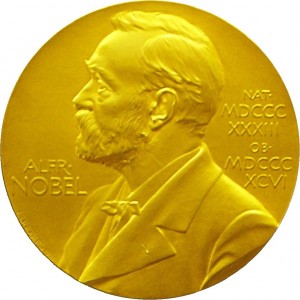
The Nobel prizes were awarded this week. Each year there are three science related awards in the fields of medicine, physics and chemistry.
In the field of medicine, the award went to John O´Keefe, May-Britt Moser and Edvard I. Moser for discovering the brain cells that make up our positioning system. In 1971 John O’Keefe discovered that when a rat was in a certain part of the room, one part of the hippocampus was always activated. When the rat was in other parts of a room there were different cells activated. He termed these cells “place cells” and determined that they formed a map. In 2005, the Mosers discovered what they called “grid cells”. These cells generated a coordinate system and aid in finding our way along paths. Read more about the physiology and medicine prize here.
This years physics medal went to the invention of LEDs and was awarded to Isamu Akasaki, Hiroshi Amano, and Shuji Nakamura. The three researchers contributed to the development of LED technology, which is prevalent in today’s telephones, lamps, and computers. LED lights emit brighter light than incandescent lights and for longer periods of time. Read more about the award at Scientific American. The press release is here.
The chemistry prize was awarded to Eric Betzig, Stefan Hell, and William Moerner for developing super resolved fluorescence microscopy. Researchers thought they were limited by the limit of diffraction when it came to resolving images under a microscope. The three Nobel recipients have developed technology that helped overcome this limitation and resolve images into the nanometer scale. Stefan Hell developed a technique called stimulated emission depletion microscopy or STED. Bezig and Moerner, working separately, performed the groundwork for the development of single molecule microscopy. You can read the press release here, and a more detailed description of high resolution microscopy here.

