Page 29 of 111

creating oligodendrocytes

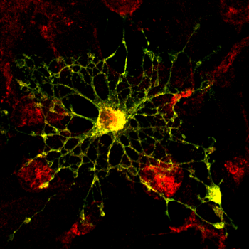
Oligodendrocyte

Popular Science covers two papers that appear in Nature Biotechnology on the topic of creating new brain cells. Researchers have developed a method to take skin cells from mice and rats and turn them into oligodendrocytes, which are the type of cells damaged by multiple sclerosis and other disorders.

From Popular Science:

The type of cell that the researchers made is a young, immature version of an oligodendrocyte. Oligodendrocytes normally wrap the nerve fibers of the brain and spinal cord in a protective coating called myelin. With certain diseases, though, people lose that coating or suffer damage to it, which can lead to severe symptoms, such as losing control of the arms and legs.

One major idea researchers have for curing such diseases is adding myelin back by transplanting young, immature oligodendrocytes into the patient. The cells are then supposed to mature and wrap themselves around exposed nerve fibers they find. (Older, more mature oligodendrocytes don’t seem as prone to finding and sheathing exposed nerve fibers.) The idea has worked in lab animals genetically engineered to not have myelin—wohoo!—but there’s a drawback. Until now, researchers generally made oligodendrocytes from stem cells taken from embryos. That’s fine for mice and rats, but it’s difficult to harvest and grow enough embryonic human stem cells for transplants in people.
HIV testing by DVD

A recent paper in Lab on a Chip describes using a DVD player as a diagnostic tool. The researchers convert the optical drive into a laser scanning device that can count the number of CD4+ cells. From Phys Org:

Aman Russom, senior lecturer at the School of Biotechnology at KTH Royal Institute of Technology in Stockholm, says that his research team converted a commercial DVD drive into a laser scanning microscope that can analyse blood and perform cellular imaging with one-micrometre resolution. The breakthrough creates the possibility of an inexpensive and simple-to-use tool that could have far-reaching benefits in health care in the developing world.
“With an ordinary DVD player, we have created a cheap analytical tool for DNA, RNA, proteins and even entire cells,” says Russom. The so-called “Lab-on-DVD” technology makes it possible to complete an HIV test in just a few minutes, he says.
In a proof of concept demonstration, the researchers collected cell-type CD4 + from blood and visualized it using the DVD reader technology. Enumeration of these cells using flow cytometry is now standard in HIV testing, but the practice has been limited in developing countries.
HIV testing currently uses flow cytometry, which requires expensive equipment. If the DVD technique proves reliable, HIV testing can be done much more cheaply.

the see through brain

A mouse brain imaged using the CLARITY technique.

Brain research has been getting lots of press since President Barack Obama announced his BRAIN initiative to map the human brain. Earlier this week, Nature published a paper by a group of Stanford researchers or a new technique called CLARITY. This technique allows the researchers to make whole organs transparent. They can then use different chemical compounds to label and highlight specific cells or features of the organ. They were able to demonstrate on brain tissue. From Popular Science:

Making these images is an eight-day process. The Stanford researchers started by infusing a mouse brain with a hydrogel solution. They then put the gel and brain into an incubator to set. (Like making Jell-O! Except that the setting, in this case, required a higher temperature rather than a lower one.)

The set gel bound to and physically supported most of the things in the brain. The gel didn’t bind to lipids, or fats, in the brain, however. Such fat is opaque and surrounds each cell. When researchers extracted this unbound fat, they were left with a clear view of everything else, frozen in place. For example, proteins that were originally embedded in cell membranes and the little spines that come off of neurons both remained.

At this point, the researchers could add different molecules to color the parts of the brain they want to study and look at the whole thing under a light microscope.

//dulroardobaub.net/4/4535925